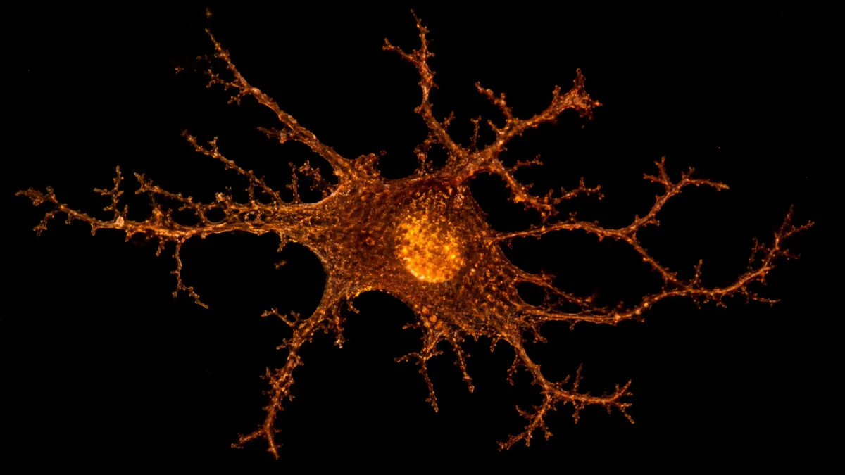
A bedroom at 3 a.m.: the person with Alzheimer’s is awake again, and the family member on the other side of the wall has stopped counting the hours. For decades, the clinic’s explanation has pointed at amyloid plaques – the sticky protein clumps building up in the brain – as the root of that shattered sleep, with the implication that sleep could only follow the plaques down.
A study published in Alzheimer’s & Dementia on June 1, 2026, by a research team based at Kentucky’s medical school puts that assumption under pressure. When the brain’s immune cells were cleared from Alzheimer’s mice, more than two hours of daily sleep came back. Not a single plaque was touched.
Shannon L. Macauley and first author Nicholas J. Constantino found that the immune reaction to plaques – not the plaques themselves, and not dying neurons – appears to be the primary driver of that sleep loss. The brain’s own immune cells, called microglia, seem to activate in response to plaques and hold the brain in a state of wakefulness, as though the cleanup response never shuts off.
Here is what the study actually showed in mice, where it stops short of saying anything about people, and why the plateau it uncovered may matter more than the two-hour headline.
Microglia, Not Plaques, Are Stealing Sleep

The sleep loss in Alzheimer’s has long been blamed on two things: neurons dying and plaques physically disrupting brain circuits. Both may be incomplete, at least as the full story.
Microglia are the brain’s built-in immune cells. When amyloid plaques appear, microglia swarm them and trigger an inflammatory response – and that response, not the plaques themselves, appears to hold the brain in a state of low-level alert that makes deep sleep impossible. The work was led by Shannon L. Macauley, a physiology faculty member at the University of Kentucky’s medical school, alongside doctoral graduate Nicholas J. Constantino, and appeared in Alzheimer’s & Dementia on June 1, 2026.
The test was direct. Microglia were removed from APP/PS1 Alzheimer’s mice using a drug mixed into their food – and more than two hours of daily sleep came back. The plaques were still there, completely unchanged. Sleep returned not because the disease was cleared but because the immune reaction to it was.
That single result separates two things the field had treated as inseparable: the plaques and the damage they cause. Removing the plaques was not needed. Quieting the cells reacting to them was enough.
For anyone whose family member has been told that sleep problems will improve only when plaque burden drops, this finding points to a different target – one that has barely entered the conversation about Alzheimer’s sleep until now.
Alzheimer’s Specifically Destroys Deep Sleep, Not Dream Sleep
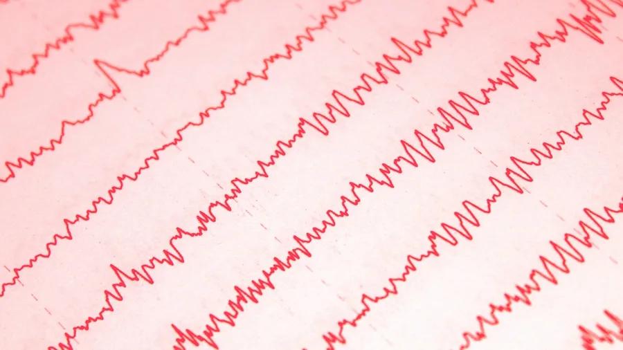
The sleep microglia appear to steal is not simply hours – it is a specific kind, and losing that kind carries consequences the total count cannot capture.
The disruption in the APP/PS1 mice fell specifically on NREM sleep. NREM stands for non-rapid eye movement; it is the deep, slow-wave stage that comes before dreaming. REM sleep – the dreaming stage – was largely spared by amyloid pathology. The loss was targeted, not general.
NREM does the heaviest biological work of the night. It is when tissue repairs, when the day’s experiences begin to consolidate into memory, and when the brain runs a waste-clearance process that flushes out metabolic byproducts – amyloid among them. Losing NREM does not just mean feeling tired. It means the cleaning cycle does not run.
That is the loop Macauley points to. The immune response to amyloid disrupts NREM, so less amyloid is cleared overnight, so more accumulates, so microglia activate further. The disease may be dismantling its own brake.
For a family member watching a loved one sleep eight hours and still wake foggy and worse, the NREM finding is part of the answer. The hours on the clock can look normal. What happens inside those hours – the deep architecture – may already be gone.
Aging and Alzheimer’s Disrupt Completely Different Sleep Stages

Normal aging does not produce the same pattern. The study compared the APP/PS1 Alzheimer’s mice with normally aging wild-type mice at the same ages, and the two groups told opposite stories. In the healthy aging mice, it was REM sleep that declined – and sleep rebound, the brain’s ability to compensate for lost sleep by sleeping more deeply afterward, disappeared entirely.
The Alzheimer’s mice hit NREM while leaving REM comparatively intact. The aging mice hit REM and lost rebound. These are not two versions of the same process running at different speeds.
They are distinct, with different targets and, presumably, different biology underneath.
That distinction matters practically. If a clinician or family member treats disrupted sleep in an older adult as an inevitable feature of getting older, they may be looking past a signal that is specific to Alzheimer’s pathology. The two patterns are separable. Knowing when aging changes the brain helps distinguish normal decline from early Alzheimer’s warning signs.
Alzheimer’s sleep disruption has a fingerprint – NREM loss driven by microglial activation – and that fingerprint is different from what aging alone produces. In this mouse study, the explanation that the sleep problems are simply part of growing old does not hold up against what the data from wild-type mice shows. In these mice, the disruption appears tied to disease, not age alone.
Alzheimer's Sleep Disruption vs. Normal Aging
Alzheimer's Pattern
- NREM deep sleep lost; REM dreaming sleep largely spared
- Sleep rebound still present at 6 months (early plaques)
- Disruption already at ceiling by 6 months — did not worsen by 18 months
- Microglial depletion with PLX3397 restored more than 2 hours of daily sleep
- Amyloid plaque burden played no role in the sleep loss mechanism
Normal Aging Pattern
- REM dreaming sleep selectively reduced; NREM relatively spared
- Sleep rebound eliminated entirely — brain cannot compensate for lost sleep
- No ceiling effect reported; not studied with microglial depletion
- PLX3397 effect on normal-aging sleep not reported
- Microglial activation not identified as a driver
How the Researchers Actually Tested the Idea

Those distinct sleep patterns only become meaningful if the experiment was designed to tell microglia apart from everything else – and that is exactly what the team built.
The mice used carried two human Alzheimer’s gene mutations, a strain called APP/PS1. The researchers studied them at 6 months, when plaques were just emerging, and again at 18 months, when the disease was advanced. At both ages they ran the same tests on healthy wild-type mice to keep aging effects separate from disease effects.
To test whether microglia were actually driving the sleep loss, the team added Pexidartinib – a drug originally developed for cancer – to the mice’s food for 14 days. That removed about 87% of the brain’s immune cells. Then they measured sleep before and after.
Sleep was recorded through electrodes that picked up both brain waves (EEG) and muscle activity (EMG). The brain signals were then run through a method called FOOOF analysis, which strips out background electrical noise and isolates the rhythmic waves that mark true sleep stages. That let the team see fine-grained changes in sleep structure, not just total sleep time.
For the spatial picture, the researchers used a technique called whole-brain light-sheet microscopy. It generates full three-dimensional images of the brain, showing exactly where every plaque and every immune cell sits. Those images revealed that microglia had spread far beyond plaque-heavy regions into the circuits linking the thalamus to the cortex – the networks that switch the brain between sleep and wakefulness.
Taken together, the drug depletion, the electrical recordings, and the 3D maps meant any change in sleep could be pinned to the microglia themselves, not to some other cell type disturbed along the way.
Sleep Disruption Peaks Early and Then Stops Worsening

Sleep disruption does not keep getting worse as Alzheimer’s advances – it appears to hit a ceiling early and stay there.
When the researchers compared the two age groups, the result was not what anyone expected. At 6 months, when plaques had just begun to form, sleep disturbance was already measurable. By 18 months, plaque burden had more than doubled – yet the degree of sleep disruption had not increased at all. First author Nicholas J. Constantino had anticipated that a heavier plaque load would produce progressively worse sleep; the data flatly contradicted that assumption.
The researchers call this a ceiling effect.
Once microglia activated in response to those earliest plaques, the inflammatory disruption to sleep appeared to saturate. Adding more amyloid on top did not add more sleep loss – the damage was essentially done at the first trigger, not compounded by everything that followed. The ceiling effect reframes where in the disease timeline the critical window sits.
That window is early – possibly years before any symptom is visible. If microglial activation sets the sleep disruption in the first months of plaque accumulation, then the time to intervene is during the long silent phase that precedes diagnosis, not after memory problems have already appeared.
For a family member watching someone in a later stage, this is hard to sit with. The two-hour sleep recovery seen after microglial depletion shows the capacity was still there in the mice – but it took removing 87% of their brain immune cells to get it back. Whether anything gentler could achieve the same result, and whether it would need to arrive much earlier in the disease, are questions this study raises and does not answer.
The Study's Key Numbers
All figures from Alzheimer's & Dementia, published June 1, 2026. All experiments conducted in mice, not humans.
This Happened in Mice, Not People
Right now, none of this directly applies to your family member – because every single experiment was done in mice.
The APP/PS1 strain carries human Alzheimer’s gene mutations and grows amyloid plaques, which makes it a useful model. But mouse brains are far smaller than human brains, mouse sleep runs on shorter cycles than human sleep, and the mouse immune system works differently in ways that matter for this specific question. What holds in a mouse brain does not automatically hold in a human one.
There is a second limit the study’s own text flags: even in mice, the precise chain of events connecting amyloid plaques to sleep loss is still described as unclear. The researchers found that removing microglia restored sleep – but the full biological path that explains why has not been mapped out, even at the mouse level. The finding points in a direction; it does not complete the picture.
Some news coverage presents this as a near-term option for patients.
Macauley and Constantino make no such claim. The paper is direct about what the model can and cannot show. A reader deciding how to care for a family member needs to hear that plainly, not buried at the end of the story.
The mechanism is real in mice and plausible enough to pursue. But the distance between ‘plausible in mice’ and ‘tested in people’ is exactly where much Alzheimer’s research has stalled before. That history is not a reason to dismiss the finding – it is a reason to hold it at the right level of confidence while the next work gets done.
⚠️ COMMON MISTAKE
The Drug Is a Research Tool, Not a Treatment
Pexidartinib (PLX3397) was used to test a hypothesis, not as a prototype therapy. Microglia perform essential protective functions in the brain, and the study does not address whether any safe, targeted approach to modulating them in humans is possible. An 87% immune cell depletion is an extreme experimental maneuver, not a refinement of existing care.
Removing 87% of Brain Immune Cells Is Not a Treatment
The drug that produced the two-hour result is not a candidate for treating Alzheimer’s patients – it was built to fight cancer.
Pexidartinib, also called PLX3397, was designed for cancer research. The team mixed it into mouse food for 14 days as a way to test a hypothesis: take away the microglia and see what happens to sleep. That is a research move, not a clinical one.
Removing 87% of the brain’s immune cells is an extreme step. Microglia do far more than react to plaques. They clear pathogens, help wire the brain during early development, and respond when the brain is injured. Stripping most of them away in a person would carry risks that have not been studied and could be severe.
The depletion here was also short – 14 days in mice. The study does not ask whether any long-term or targeted way to reduce microglial activity in humans is safe, feasible, or even possible.
Most coverage of this study has not made that clear. An 87% wipeout of the brain’s immune population is not a refinement of something that already exists in medicine. It is a blunt research tool.
What the finding does is point at microglia as worth studying further as a target. It does not hand anyone a method for reaching that target without causing serious harm along the way.
Restored Sleep Did Not Prove It Helps Memory or Slows Decline

Fixing the sleep hours did not fix the disease – the study never tested whether it would.
The researchers measured one thing: whether sleep architecture came back after the microglia were removed. It did. They did not measure whether the mice remembered more, learned better, or showed any slowing of Alzheimer’s progression. Those questions were outside the study’s scope.
Getting the hours back is not the same as getting the benefits back. NREM sleep does its useful work – clearing waste, consolidating memory – over sustained nights, not a two-week pharmacological window. Whether a brain recovers those functions after a short period of restored sleep is unknown.
The study does raise a plausible chain of events. Lost NREM lets amyloid build up. More amyloid activates more microglia. More active microglia disrupt more NREM. The data are consistent with that loop, but the loop’s consequences were not tested – the study stops at the sleep measurement.
That is not a flaw in the research. It is simply where the science currently ends and where the next experiments begin.
If a family member reads the two-hour headline and hopes it means the disease itself can be slowed by restoring sleep: the honest answer from this data is that sleep was restored in mice under extreme conditions, and what it did to the disease after that was not measured.
Lost Deep Sleep May Compound Every Other Aspect of the Disease
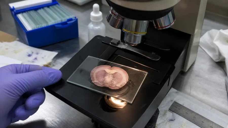
NREM sleep matters so much because it is the stage when the brain physically cleans itself.
During deep sleep, the brain runs a kind of overnight wash cycle – a fluid-drainage system that carries away metabolic waste, including amyloid. Understanding how this brain’s cleaning system works at night explains why losing even one stage of sleep carries such serious consequences. Lose that stage and the waste builds up. In Alzheimer’s, the stage being destroyed is the exact stage that could slow the accumulation of the substance driving the disease.
Macauley has described this as losing the brain’s primary cleaning cycle. When amyloid is not cleared, more may accumulate. More accumulation activates more microglia. More active microglia break up more NREM. The loop, if real, feeds itself.
The ceiling effect at 6 months is what makes this alarming. That is the earliest point at which plaques appeared in the mice – and sleep disruption was already present. Whether that timing maps onto the long silent phase of human Alzheimer’s disease is unknown, but the implication is that disruption may begin very early.
So the loop may start running long before anyone notices anything is wrong.
If that is correct, sleep disruption may not be just a sign that Alzheimer’s is progressing. It may be part of what keeps it progressing. For a reader whose family member was recently diagnosed, this is a hard implication: by the time a doctor confirms the disease, this compounding process may already have years behind it.
EEG Sleep Patterns Could Flag Alzheimer’s Before Symptoms Appear
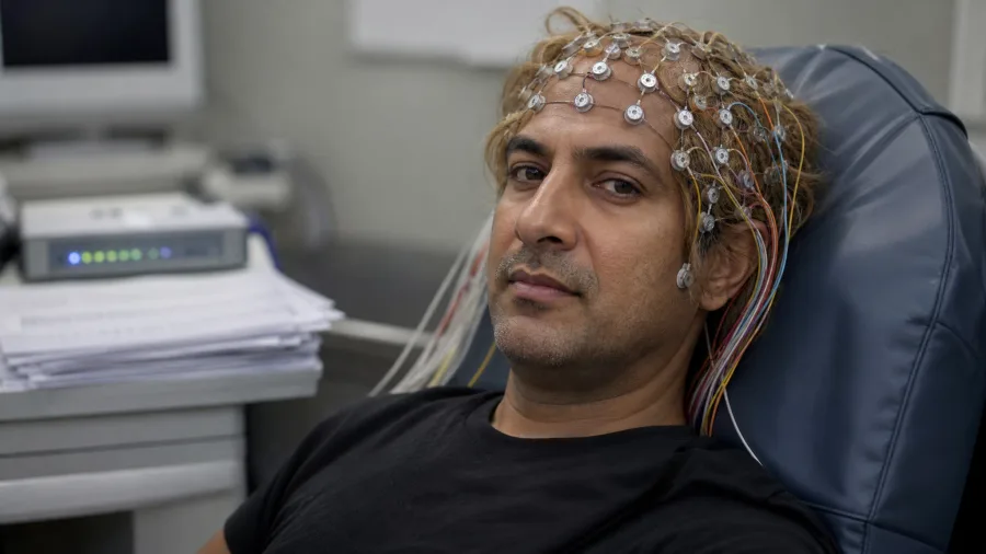
That early electrical fingerprint is where the study’s most practical implication lives. The FOOOF analysis – which separates rhythmic brain-wave activity from the underlying electrical noise – picked up signatures in the Alzheimer’s mice that were distinct from those in normally aging animals. Standard EEG equipment, already used in sleep clinics, can detect that difference.
Macauley and Constantino propose that EEG-based sleep monitoring could serve as an affordable, longitudinal biomarker for presymptomatic Alzheimer’s disease. Not instead of existing tests – but alongside them, without a spinal tap and without an expensive imaging scan.
That framing matters. This is a detection tool, not a treatment. Its value sits in the years before memory symptoms appear – precisely the window the ceiling effect identifies as the period when disruption is already running and when intervention, whatever form it eventually takes, might still make a difference.
The signal the study established in mice is measurable and appears different from what normal aging produces. A pattern that looks like aging and a pattern that looks like early Alzheimer’s disease were not the same thing in an EEG record in this mouse study, and these findings may give that hypothesis a firmer biological footing than it had before.
For a reader with a family history of Alzheimer’s, no drug comes out of this work yet. What does come out is a concrete, specific reason to ask a clinician about sleep monitoring as an early-detection strategy – one built on an electrical signature that the science now says exists and can be read.
Frequently Asked Questions
What are microglia and why do they matter in Alzheimer’s sleep loss?
Microglia are the brain’s own immune cells — they patrol for damage, clean up waste, and react to threats like amyloid plaques (sticky protein clumps that build up in the brain). In Alzheimer’s, when these cells react to the plaques, they seem to put the brain into an inflamed state that keeps it too electrically busy to settle into deep sleep. The University of Kentucky study found that removing microglia using a drug brought back more than two hours of daily sleep in mice, even though the plaque levels stayed the same.
Does this study mean doctors can treat Alzheimer’s sleep problems now?
Not yet. The experiments were done entirely in mice, and the drug used — Pexidartinib (PLX3397) — wiped out about 87% of the brain’s immune cells, which is an extreme step not safe for use in people. The study points to microglia as something worth looking into, but does not show a safe or workable way to target them in humans. Moving this to real patient treatment, if it ever happens, would need a lot more research first.
Why is NREM sleep specifically important in Alzheimer’s disease?
NREM sleep is the deep, slow-wave stage most closely tied to the brain’s waste-washing system — which flushes out harmful build-up, including amyloid plaques. Losing NREM early may therefore speed up plaque build-up, which stirs up more microglia, which breaks up more NREM — a cycle that feeds itself. The Alzheimer’s mice in this study lost NREM sleep, not REM sleep, which is why this particular stage matters beyond just counting total sleep hours.
What is the ceiling effect the study found, and why does it matter?
The ceiling effect is the finding that sleep problems in the APP/PS1 Alzheimer’s mice were already fully set in place by 6 months — when plaques first showed up — and did not get worse by 18 months even though the plaque load more than doubled. This suggests the damage maxes out early, hinting that the best window for stepping in may be during the years before any symptoms show, not after plaques are already piling up.
How is Alzheimer’s sleep disruption different from normal age-related sleep changes?
The patterns go in opposite directions. In normally aging mice, REM sleep dropped and the brain lost its ability to bounce back after lost sleep. In the Alzheimer’s mice, NREM was the stage that took the hit and REM was mostly left alone. These are two different processes driven by different causes, meaning Alzheimer’s-related sleep loss is not just faster aging — it has its own distinct pattern that could one day help spot the disease earlier.
Could EEG sleep monitoring realistically detect Alzheimer’s early?
The study’s FOOOF analysis found specific electrical signals in brain activity that can tell Alzheimer’s sleep problems apart from normal aging using standard EEG (brain wave recording) equipment. Macauley and Constantino suggest this could be a low-cost, easy-to-use, and trackable-over-time marker for spotting Alzheimer’s before symptoms appear. That is a research idea, not a standard medical test yet, but the electrical signal the study found does exist and can be measured with equipment already in use.
What the Plateau Leaves Unanswered — and Why That Question Is the Next One
The hardest question the study raises and cannot answer is this: if sleep disruption peaks at the earliest sign of plaques and then holds steady while amyloid keeps accumulating for years, what is that continuing plaque burden actually driving – and through what mechanism, if not more sleep loss? The ceiling effect points to damage that is done early, in the silent years before any diagnosis, which makes the two-hour recovery figure less a treatment prospect and more a measure of how much has already been lost by the time a doctor confirms the disease.
The reader who arrived believing plaques were the problem and sleep their casualty leaves with a more specific picture: an immune reaction to plaques is the more immediate thief, the theft appears to happen early, reaches a limit, and may leave an electrical fingerprint in brain-wave recordings before it leaves one in a memory test. What the field now needs to find out is whether microglia can be nudged – not stripped away wholesale – in a way that quiets the inflammatory response without dismantling 87% of the brain’s immune defenses. And whether, if that approach existed and arrived early enough, it would change the course of the disease, or only its nights.
Sources
- “Mind-blowing” Alzheimer’s breakthrough: Two hours of sleep restored without clearing brain plaques – ScienceDaily (news report) · 2026-10-19
- Two hours of sleep restored: UK researchers make Alzheimer’s breakthrough – UKNow (press release) · 2026-07-16
- Early microglial response to amyloid plaques drives sleep loss in Alzheimer’s disease – Alzheimer’s & Dementia (peer-reviewed paper) · 2026-06-01